
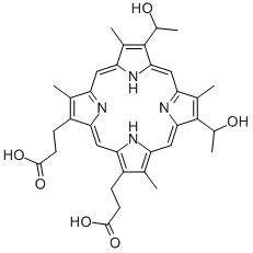
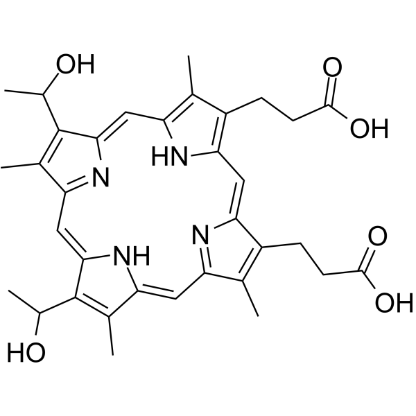
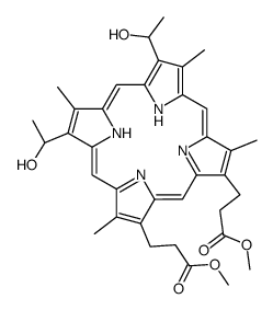
Hematoporphyrin 血卟啉; Hematoporphyrin IX,95.81%
产品编号:Bellancom-B0754| CAS NO:14459-29-1| 分子式:C34H38N4O6| 分子量:598.69
Hematoporphyrin (Hematoporphyrin IX) 是血红素结合蛋白亲和色谱底物。
本网站销售的所有产品仅用于工业应用或者科学研究等非医疗目的,不可用于人类或动物的临床诊断或者治疗,非药用,非食用,
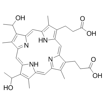
Hematoporphyrin 血卟啉; Hematoporphyrin IX
| 产品介绍 | Hematoporphyrin (Hematoporphyrin IX) 是一种光敏剂,是血红素结合蛋白亲和色谱底物。当暴露于红光下时,Hematoporphyrin 可以诱导 U87 胶质瘤细胞凋亡,并降低体内肿瘤的生长。 | ||||||||||||||||
|---|---|---|---|---|---|---|---|---|---|---|---|---|---|---|---|---|---|
| 生物活性 | Hematoporphyrin (Hematoporphyrin IX), a photosensitizer, is a substrate for affinity chromatography of heme-binding proteins. Hematoporphyrin can induce apoptosis in U87 glioma cells and decrease tumor growth in vivo when exposed to red light. | ||||||||||||||||
| 体外研究 |
Hematoporphyrin (20-120 nM; 60 min) dose-dependently inhibits cell viability in U87 and U251 glioma cells, with IC50s of 85 and 166 nM, respectively. 西域 has not independently confirmed the accuracy of these methods. They are for reference only. Cell Viability Assay
Apoptosis Analysis
|
||||||||||||||||
| 体内研究 (In Vivo) |
Hematoporphyrin (5-10 mg/kg; i.p. for 2 months) with the irradiation of red light rapidly decreases the tumor size of rats, due to necrosis caused both by direct action of the photoactivated porphyrin on the tumor cells and by secondary effects on blood vessels. 西域 has not independently confirmed the accuracy of these methods. They are for reference only.
| ||||||||||||||||
| 体内研究 |
Hematoporphyrin (5-10 mg/kg; i.p. for 2 months) with the irradiation of red light rapidly decreases the tumor size of rats, due to necrosis caused both by direct action of the photoactivated porphyrin on the tumor cells and by secondary effects on blood vessels. 西域 has not independently confirmed the accuracy of these methods. They are for reference only.
| ||||||||||||||||
| 性状 | Solid | ||||||||||||||||
| 溶解性数据 |
In Vitro:
DMSO : 150 mg/mL (250.55 mM; Need ultrasonic) 配制储备液
*
请根据产品在不同溶剂中的溶解度,选择合适的溶剂配制储备液;该产品在溶液状态不稳定,建议您现用现配,即刻使用。 In Vivo:
请根据您的实验动物和给药方式选择适当的溶解方案。以下溶解方案都请先按照 In Vitro 方式配制澄清的储备液,再依次添加助溶剂:
——为保证实验结果的可靠性,澄清的储备液可以根据储存条件,适当保存;体内实验的工作液,建议您现用现配,当天使用;
以下溶剂前显示的百
| ||||||||||||||||
| 运输条件 | Room temperature in continental US; may vary elsewhere. | ||||||||||||||||
| 储存方式 |
-20°C, protect from light *该产品在溶液状态不稳定,建议您现用现配,即刻使用。 | ||||||||||||||||
| 参考文献 |
|
| 安全声明 (欧洲) | 22-24/25 |
|---|---|
| WGK德国 | 3 |
| RTECS号 | TS5500000 |
|
~%
14459-29-1 |
| 文献:Australian Journal of Chemistry, , vol. 33, # 3 p. 585 - 597 |
|
~%
14459-29-1 |
| 文献:Hoppe-Seyler's Zeitschrift fuer Physiologische Chemie, , vol. 142, p. 141,151, 145<1925>202,214 |
|
~%
14459-29-1 |
| 文献:Justus Liebigs Annalen der Chemie, , vol. 468, p. 98,108 |
|
~%
14459-29-1 |
| 文献:Justus Liebigs Annalen der Chemie, , vol. 468, p. 98,108 |
|
~%
14459-29-1 |
| 文献:Australian Journal of Chemistry, , vol. 33, # 3 p. 585 - 597 |
 有竞争力的价格
匹配竞争对手的价格
 极速物流
极速物流效率为先
 技术支持
技术支持专业经验 贴心服务
 现货库存
现货库存50000+库存

 浙公网安备 33010802013016号