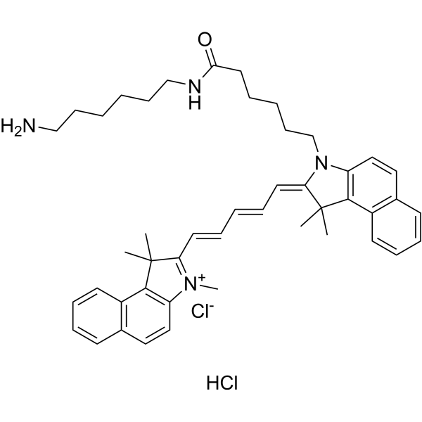
Cyanine5.5 amine Cy 5.5 amine; Lumiprobe Cy 5.5 amine,98.23%

产品编号:Bellancom-D1540| CAS NO:2097714-45-7| 分子式:C46H58Cl2N4O| 分子量:753.88

本网站销售的所有产品仅用于工业应用或者科学研究等非医疗目的,不可用于人类或动物的临床诊断或者治疗,非药用,非食用,

货号 包装 价格 库存与货期 购买量 操作
Bellancom-D1540
1100.00 杭州 北京(现货)
Bellancom-D1540
2200.00 杭州 北京(现货)
Bellancom-D1540
3080.00 杭州 北京(现货)

增值税发票√顺丰快递√订货电话:18601927057

Cyanine5.5 amine Cy 5.5 amine; Lumiprobe Cy 5.5 amine

产品介绍 Cyanine5.5 amine (Cy 5.5 amine),Cy5.5 类似物,是一种近红外 (NIR) 荧光染料 (Ex=648 nm,Em=710 nm)。Cyanine5.5 amine 可用于制备 Cy5.5 标记的纳米颗粒,从而得以用共聚焦显微镜进行跟踪和成像,且荧光背景低。
生物活性

Cyanine5.5 amine (Cy 5.5 amine), a Cy5.5 Analogue, is a near-infrared (NIR) fluorescent dye (Ex=648 nm, Em=710 nm). Cyanine5.5 amine can be used in the preparation of Cy5.5-labeled nanoparticles, which can be tracked and imaged with low fluorescence background using confocal microscopy.

体外研究
体内研究

Real-time monitoring Cy5.5-labeled nanoparticles (Cy5.5-PLGA) in retinal blood vessels.
Guidelines (Following is our recommended protocol. This protocol only provides a guideline, and should be modified according to your specific needs).
1. Freshly prepared suspensions of Cy5.5-PLGA (resuspended in 0.5 ml 1% poloxamer 188, vortexed gently and left for 30 min at ambient temperature before use) is administrated intravenously (0.5 mL).
2. Anesthetize the rats, treat the eyes with Neosynephrine-POS 5% to relax the iris, and Vidisic eye gel is applied to protect the eye from drying out and used as immersion medium for the contact lens as well.
3. Fix the rats under a confocal scanning microscope with the eye positioned in working distance underneath the objective lens, and a cannula is inserted into the tail vein.
4. Observe the fluorescence in the retina, and capture the images at different time points (0, 1, 3, 5, 15, 30, 60, 90 min).
Note: The rats are kept on the heating plate during all the in vivo imaging process.
5. After in vivo real-time imaging, rats were euthanized with an overdose of aforementioned anesthetic and the eyeballs were enucleated and placed into cooled HEPES buffered solution (135 mM NaCl, 5 mM NaOH, 2.5 mM KCl, 7 mM MgCl2, 10 mM HEPES, 10 mM glucose; pH7.4).
6. Remove the anterior segment of eye and vitreous body, separate whole retina carefully, flat on the modified culture plate.
7. Incubate the whole mounts with0.1 mg/mL Hoechst 33342 (HY-15559) in HEPES solution for 20 min for nuclei staining.
8. Fix flat mount retina with 4% paraformaldehyde solution for 20 min and wash with HEPES solution.
9. Capture the images immediately with microscope after preparation of retinal flat mount.

西域 has not independently confirmed the accuracy of these methods. They are for reference only.

性状Solid
溶解性数据
In Vitro: 

DMSO : 125 mg/mL (165.81 mM; ultrasonic and warming and heat to 60°C)

配制储备液
浓度 溶剂体积 质量 1 mg 5 mg 10 mg
1 mM 1.3265 mL 6.6324 mL 13.2647 mL
5 mM 0.2653 mL 1.3265 mL 2.6529 mL
10 mM 0.1326 mL 0.6632 mL 1.3265 mL
*

请根据产品在不同溶剂中的溶解度选择合适的溶剂配制储备液;一旦配成溶液,请分装保存,避免反复冻融造成的产品失效
储备液的保存方式和期限:-80°C, 6 months; -20°C, 1 month (protect from light)。-80°C 储存时,请在 6 个月内使用,-20°C 储存时,请在 1 个月内使用。

In Vivo:

请根据您的实验动物和给药方式选择适当的溶解方案。以下溶解方案都请先按照 In Vitro 方式配制澄清的储备液,再依次添加助溶剂:

——为保证实验结果的可靠性,澄清的储备液可以根据储存条件,适当保存;体内实验的工作液,建议您现用现配,当天使用; 以下溶剂前显示的百
分比是指该溶剂在您配制终溶液中的体积占比;如在配制过程中出现沉淀、析出现象,可以通过加热和/或超声的方式助溶

  • 1.

    请依序添加每种溶剂: 10% DMSO    40% PEG300    5% Tween-80    45% saline

    Solubility: 2.08 mg/mL (2.76 mM); Suspended solution; Need ultrasonic

    此方案可获得 2.08 mg/mL (2.76 mM) 的均匀悬浊液,悬浊液可用于口服和腹腔注射。

    以 1 mL 工作液为例,取 100 μL 20.8 mg/mL 的澄清 DMSO 储备液加到 400 μL PEG300 中,混合均匀;向上述体系中加入50 μL Tween-80,混合均匀;然后继续加入 450 μL生理盐水定容至 1 mL。

    将 0.9 g 氯化钠,完全溶解于 100 mL ddH₂O 中,得到澄清透明的生理盐水溶液
  • 2.

    请依序添加每种溶剂: 10% DMSO    90% (20% SBE-β-CD in saline)

    Solubility: 2.08 mg/mL (2.76 mM); Suspended solution; Need ultrasonic

    此方案可获得 2.08 mg/mL (2.76 mM) 的均匀悬浊液,悬浊液可用于口服和腹腔注射。

    以 1 mL 工作液为例,取 100 μL 20.8 mg/mL 的澄清 DMSO 储备液加到 900 μL 20% 的 SBE-β-CD 生理盐水水溶液中,混合均匀。

    将 2 g 磺丁基醚 β-环糊精加入 5 mL 生理盐水中,再用生理盐水定容至 10 mL,完全溶解,澄清透明
*以上所有助溶剂都可在 西域 网站选购。
运输条件

Room temperature in continental US; may vary elsewhere.

储存方式

-20°C, protect from light

*In solvent : -80°C, 6 months; -20°C, 1 month (protect from light)

参考文献

相关文档

化学品安全说明书(MSDS)

下载MSDS

质检证书(COA)

相关产品


服务热线

13911702513
18601927057

微信客服